
Eye Site

Pterygium and Pingueculum
These degenerative conditions on the inner corner of the eye are very common in Western Australia and probably relate to sun and dust exposure.
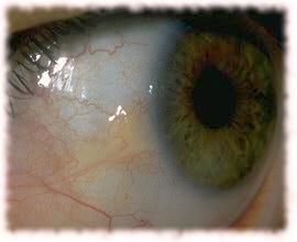 |
Pingueculum This is a yellow-white mass at the inner corner of the eye. The surface may be raised and rough causing irritation and redness. It is unlikely to cause any visual problems but may interfere with the ability to tolerate contact lenses |
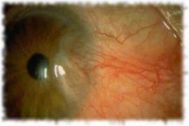 |
Pterygium This is a fleshy overgrowth of tissue spreading from the inner corner of the eye across the cornea. Initially it be similar to a Pingueculum and may cause irritation and redness. If it extends far onto the cornea it will start to reduce vision. |
The cause for these two conditions seems to be sun and dust exposure so prevention by wearing sun protection in the form of UV filter sunglasses and a hat are extremely important.
Patients present with redness , irritation and less frequently with reduction in vision. Careful analysis of the corneal surface using the Orbscan Topography Instrument will indicate if there is any distortion of the cornea occurring. If distortion or reduction of vision is occurring then surgical removal of the pterygium is required as the distortion and scarring may become permanent.
For minor problems conservative measures may be adequate with use of sun protection combined with lubricant drops to help prevent the eyelid catching on the rough surface. If the lesion is inflamed and red use of anti-inflammatory drops such as antihistamines may help. Topical steroids are usually very effective in this situation but may have very serious visual complications if used for prolonged periods. An infrequent short burst of steroid drops is relatively safe.
If the lesion is threatening the sight , or if the cosmetic appearance is unsatisfactory or if there is too much discomfort then surgical excision can be performed.
Surgery
The main problem with excision of Pterygia is that recurrence is common often happening
within a few months. In my experience of all the methods used to prevent recurrence the
most successful is conjunctival autografting. I have been performing this procedure since
1984 on recurrent pterygia and since 1988 for almost all pterygium excisions. During that
time I have seen only 2 recurrences that were clinically significant. (several minor
recurrences have been seen without any symptoms or visual effects.)
Other methods of preventing recurrence include anti-metabolites and radiotherapy both of
which are associated with a significant risk of complication at a later date.
Procedure
The operation is usually performed under local anaesthetic and can be day surgery.
After anaesthetic drops are applied a speculum is used to hold the eye open and prevent blinking.
A microscope is used and it’s light is very bright.
The pterygium is scrapped off then a partial thickness patch graft is obtained from under
the top eyelid.
This is sewn into place with sutures far finer than a hair and the eye is padded.
The whole operation takes about 20 minutes and causes little discomfort at the time (apart from the bright light). When the anaesthetic wears off there may be considerable discomfort but this can usually be controlled using a variety of medications ( see the operation information sheet )
Discomfort rapidly reduces and is usually minor by the third day and gone by one week. Most patients are able to return to work by the second or third day.